

Dental Technology in Beverly Hills
Dental Technology
3D imaging, digital scanning, soft tissue lasers and ozone, all in this office. Equipment most general practices refer out for, so planning and treatment happen in the same place.
Why the Equipment in the Room Matters
Technology in dentistry is easy to advertise and harder to justify. What it should do is make treatment more accurate, more comfortable, or possible in one place rather than three. Everything here earns its place on one of those grounds. A cone beam scan measures bone in three dimensions, so an implant is planned rather than judged. A digital scanner replaces impression trays, so restorations fit better at the margin. A soft tissue laser reshapes gum tissue with less bleeding and fewer sutures. Ozone reduces bacterial levels at a treated site as an adjunct to conventional care. None of it replaces clinical judgement. It gives that judgement better information to work with.
THE TECHNOLOGY WE USE AND WHY
Five pieces of equipment and what each one changes about the treatment you receive.

3D Cone Beam CT Scanning
A cone beam scan produces a three-dimensional image of your jaw, showing bone height and width, the position of the nerve canal, the sinus floor and the full root anatomy of a tooth. Used for implant planning, complex root canal cases and wisdom teeth. It replaces estimation with measurement.
Digital X-Rays
Digital sensors use a lower radiation dose than the film systems they replaced and produce an image on screen instantly. Decay between teeth and beneath old restorations is invisible to the eye, and this is how it is found while it is still small.
Intraoral Digital Scanner
A small wand captures your teeth as an accurate digital model, replacing impression trays and the material that sets in your mouth. Crowns, onlays, veneers, aligners and night guards are all made from that scan, and the fit at the margin is consistently better.
Soft Tissue Laser
Used to reshape gum tissue, treat the lining of periodontal pockets after cleaning, and contour the gum line before restorations. It seals small blood vessels as it works, so there is typically less bleeding, often no sutures, and a more comfortable recovery than a scalpel.
Ozone Therapy
Applied as an adjunct at a treated site, whether a prepared cavity, a cleaned periodontal pocket or an extraction socket, to reduce bacterial levels. It supports conventional treatment rather than replacing it, and we are clear about that distinction.
Planning Before Treatment, Not During
Imaging is taken and reviewed before a procedure begins, not to confirm what has already been decided. Surprises are what turn straightforward cases into difficult ones, and 3D planning removes most of them.
Fewer Appointments and Fewer Referrals
Because the imaging and the treatment happen in the same office, there is no separate scan appointment elsewhere and no waiting for images to arrive from another provider before your plan can be finalised.
Lower Radiation, Better Information
Digital imaging delivers a lower dose than film while producing images that can be enlarged, adjusted and compared against previous visits. Images are taken at intervals matched to your risk rather than automatically at every appointment.
You See What We See
Scans and X-rays appear on a screen in the room. Being shown the crack in your own tooth or the bone level around it makes treatment decisions considerably easier than being told about them.
DIAGNOSIS AND TREATMENT IN ONE PLACE
The practical benefit of having this equipment on site is not novelty. It is that fewer things are estimated and fewer things are outsourced.
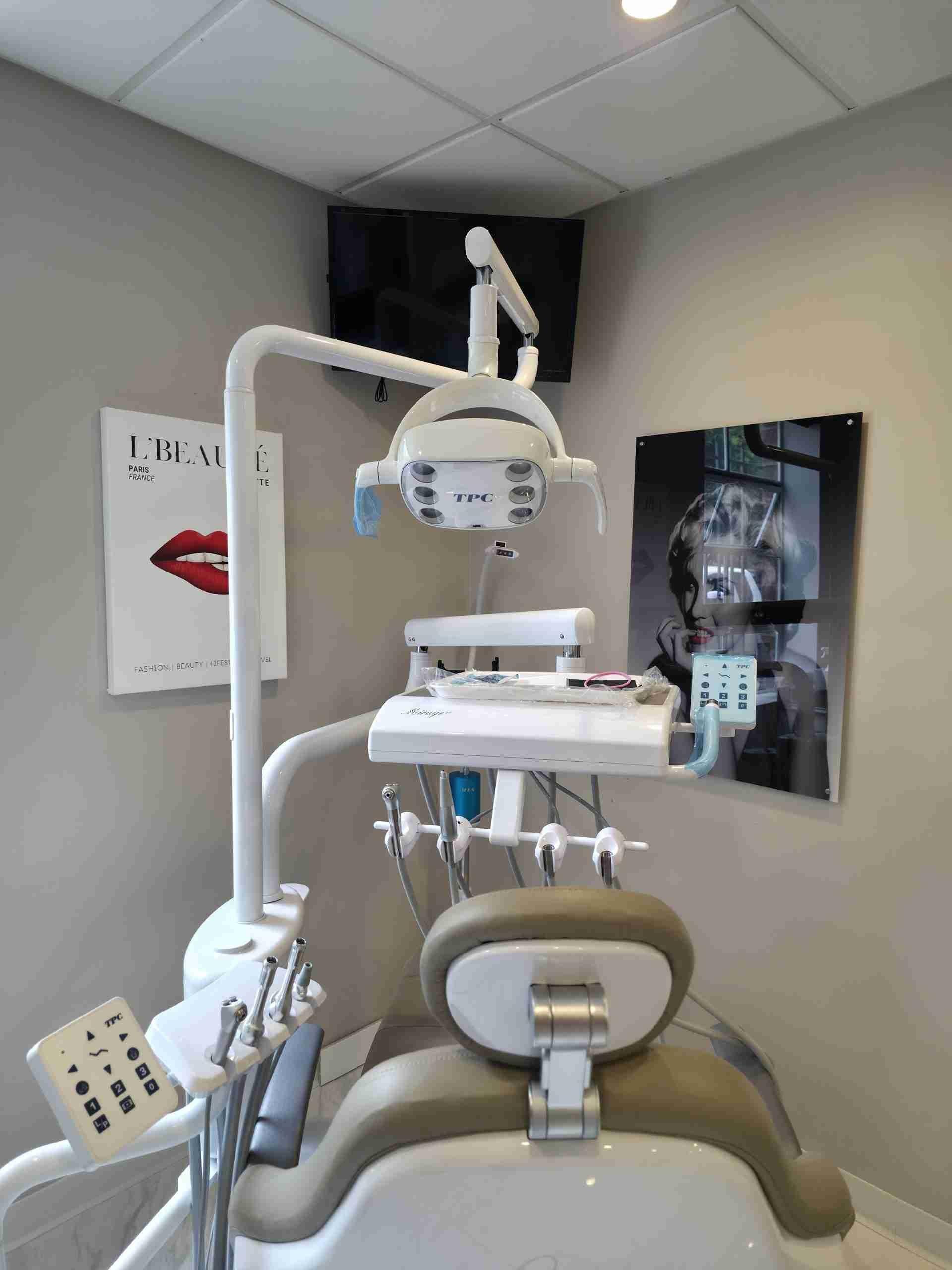
MORE ACCURATE DIAGNOSIS:
Cone beam imaging measures bone in three dimensions where a flat X-ray only approximates height. Digital X-rays find decay between teeth years before it becomes visible or uncomfortable. An intraoral camera shows you what we are describing rather than asking you to take our word for it.
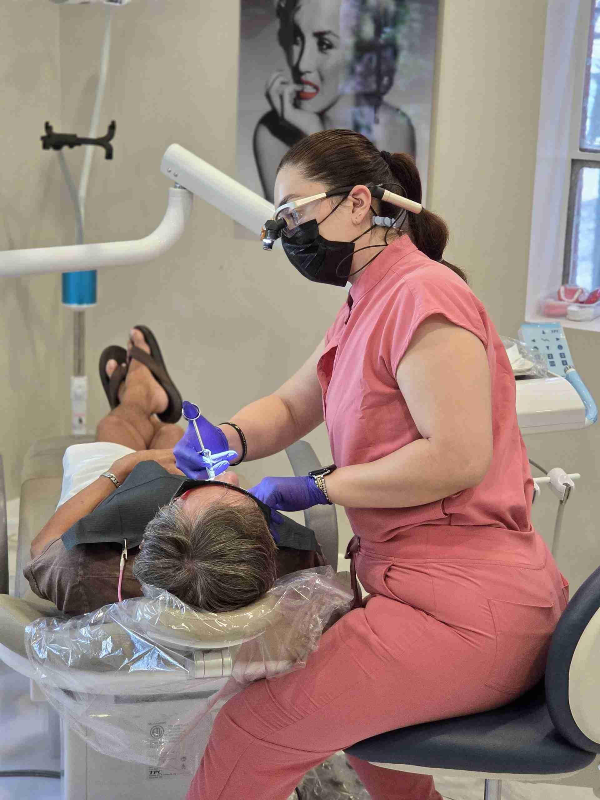
MORE COMFORTABLE TREATMENT:
Digital scanning removes impression trays and the material that sets in your mouth. Laser treatment usually means less bleeding, fewer sutures and a quicker recovery than a scalpel. Planning from a 3D scan means procedures run to a plan rather than adapting to what is found partway through.
WHY IT MATTERS THAT IT IS ALL IN ONE OFFICE
Most general practices refer out for at least some of this. Having it here changes the sequence rather than just the equipment list.

No Referrals for Imaging
Cone beam scans are taken here, at the same appointment as your assessment, rather than at a separate imaging centre with a wait before your plan can be finalised.

Planned and Treated by One Dentist
Dr. Shir reviews the imaging and carries out the treatment, so the finished result follows the plan rather than being adapted to it afterwards.

A Biological Approach
Ozone, metal-free materials and conservative preparation run through how the practice works, with technology chosen to support that rather than to replace it.

30+ Years of Experience
More than three decades of practice. Equipment is a tool, and what it produces still depends on the judgement of the person using it.
No Referrals for Imaging
Cone beam scans are taken here, at the same appointment as your assessment, rather than at a separate imaging centre with a wait before your plan can be finalised.
Planned and Treated by One Dentist
Dr. Shir reviews the imaging and carries out the treatment, so the finished result follows the plan rather than being adapted to it afterwards.
A Biological Approach
Ozone, metal-free materials and conservative preparation run through how the practice works, with technology chosen to support that rather than to replace it.
30+ Years of Experience
More than three decades of practice. Equipment is a tool, and what it produces still depends on the judgement of the person using it.
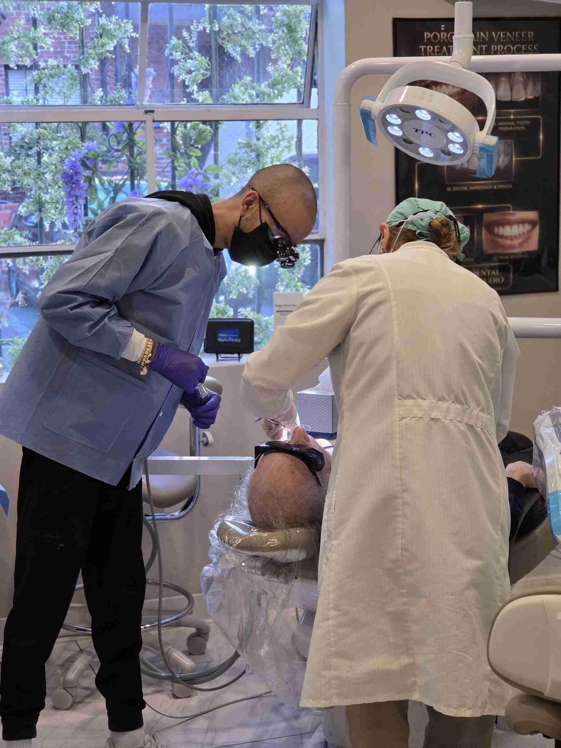
SEE YOUR OWN SCAN AT YOUR CONSULTATION
Most patients have never seen their own teeth in three dimensions, and it makes decisions about treatment considerably clearer. Whether you are considering an implant, have been told a tooth needs removing, or simply want a proper assessment of where you stand, the imaging happens here and you see it on the screen with Dr. Shir. Book a consultation.
FREQUENTLY ASKED QUESTIONS ABOUT OUR TECHNOLOGY
The questions patients ask most often about the equipment we use.
Is dental imaging safe?
Digital X-rays use a considerably lower radiation dose than the film systems they replaced, and the dose from a routine set is very small. A cone beam scan uses more than a standard X-ray but far less than a medical CT, and it is taken only where the clinical situation genuinely calls for it. Protective shielding is used, and if you are pregnant, tell us and we will discuss whether imaging should wait.
Do I still need impressions for a crown or night guard?
In most cases no. An intraoral scanner captures your teeth digitally, which means nothing sets in your mouth and no tray needs to be tolerated. The scan is also more accurate at the margin, which is where restorations succeed or fail over time. Occasionally a conventional impression is still the better option, and we will tell you if yours is one of those cases.
Why do I need a 3D scan when my old dentist only took X-rays?
A flat X-ray shows height and shape but cannot measure width, and width is frequently the dimension that determines whether an implant is possible. It also cannot show exactly where the nerve canal runs or how close a root sits to the sinus. For implants, complex root canals and wisdom teeth, that information changes the plan rather than just confirming it.
Does better technology mean higher fees?
Not in the way people expect. Having the equipment here removes the separate imaging fee you would pay elsewhere and the extra appointment that comes with it. More importantly, finding problems while they are small is what keeps treatment at the cheaper end. You receive a written figure before anything begins either way.