

3D CT Scans in Beverly Hills
Dental Technology
Cone beam imaging taken in this office. Bone measured in three dimensions, nerve position located, and root anatomy seen properly before treatment begins.
What a Flat X-Ray Cannot Show You
A standard dental X-ray is a two-dimensional image of a three-dimensional structure. It shows height and shape reasonably well, but it cannot measure width, and it flattens everything onto one plane, so a root sitting in front of the nerve canal looks identical to one sitting on top of it. Cone beam CT builds a three-dimensional model of your jaw instead. Bone height and width are measured directly, the inferior alveolar nerve is located, the sinus floor is visible, and the full root anatomy of a tooth can be seen from every angle. For implants, complex root canals and wisdom teeth, that is the difference between planning a procedure and judging it as you go.
WHERE WE USE 3D IMAGING
A cone beam scan is not taken routinely. These are the situations that genuinely call for it.

Dental Implant Planning
Bone height and width at the site are measured directly, along with the position of the nerve canal and the floor of the sinus. Width is frequently the dimension that decides whether an implant is possible, and a flat X-ray cannot measure it at all.
Second Opinions on Bone Volume
Patients told elsewhere that they lack the bone for an implant are sometimes candidates once the site is measured in three dimensions rather than estimated from a flat image. It is worth having reassessed before accepting that answer.
Wisdom Teeth Removal
Lower wisdom tooth roots often sit close to the inferior alveolar nerve. The scan shows exactly how close, which changes how the tooth is approached and is the single most useful piece of information before that procedure.
Complex Root Canal Cases
Root anatomy varies considerably between patients, and canals missed during treatment are a well-documented cause of later failure. Where a tooth has unusual anatomy or has been treated before, the scan shows the canal system in three dimensions.
Extractions Near the Sinus
Upper molar roots can sit against or through the sinus floor. Knowing that in advance changes the technique and avoids a complication that is far easier to prevent than to manage.
Airway Assessment
Where a scan has been taken for another purpose, the airway space is visible on it. That can support a conversation about sleep apnea risk, though it does not diagnose the condition, which requires a sleep study interpreted by a physician.
Jaw Joint Imaging
For TMJ cases where the picture is unclear, the joint structures can be viewed in three dimensions rather than inferred from symptoms alone.
Radiation and When We Take One
A cone beam scan uses more radiation than a standard dental X-ray and considerably less than a medical CT. It is taken only where the clinical situation genuinely calls for it, never routinely, and protective shielding is used.
Reviewed With You on Screen
The scan is rotated and sectioned on a monitor in the room. Most patients have never seen their own jaw in three dimensions, and it makes decisions about implants or extractions considerably clearer.
CONE BEAM CT AND STANDARD X-RAYS
They are not competing options. They answer different questions, and most patients only ever need the standard one.
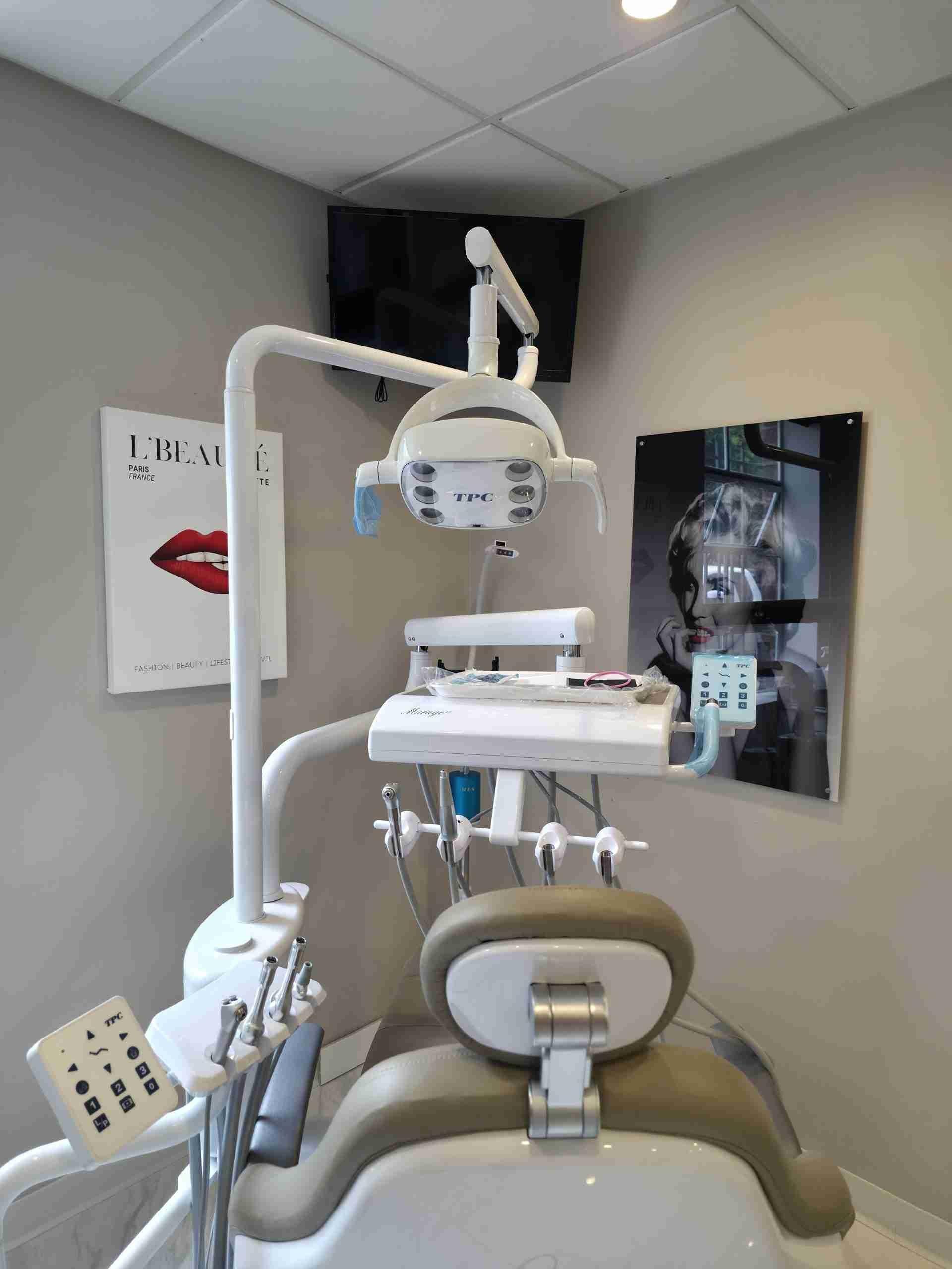
STANDARD DIGITAL X-RAYS:
The routine imaging used at examinations. They show decay between teeth, failing margins under existing fillings, bone levels and infection at root tips, at a very low radiation dose. This is what the overwhelming majority of patients need, and nothing more.
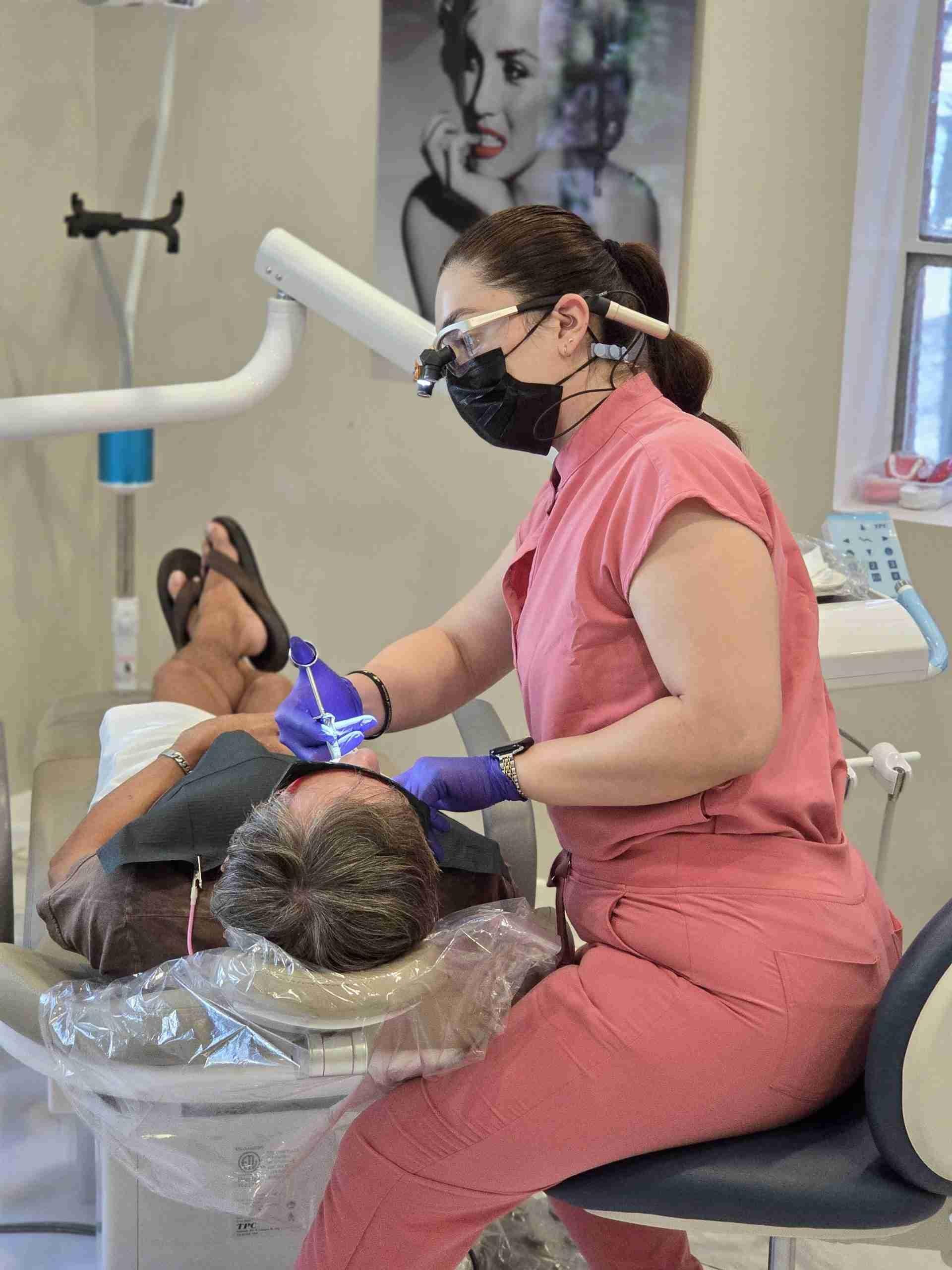
CONE BEAM CT:
Used where a two-dimensional image is genuinely not enough: implant planning, wisdom teeth close to the nerve, complex or previously treated root canals, and upper molars sitting against the sinus. Higher dose than a standard X-ray and far lower than a medical CT, and taken only when the clinical situation requires it.
WHY IT MATTERS THAT THE SCANNER IS HERE
Most general practices refer out for cone beam imaging. Having it on site changes the sequence, not just the equipment list.

No Separate Imaging Appointment
The scan is taken at your consultation rather than at an imaging centre, with no wait for files to arrive before your plan can be finalised.

Planned and Treated by One Dentist
Dr. Shir reviews the scan and carries out the treatment, so the procedure follows the plan the imaging produced rather than being adapted afterwards.

Taken Only When Indicated
A cone beam scan is never routine here. It is taken where the clinical situation genuinely calls for it, and we will explain what we are looking for.

30+ Years of Experience
More than three decades of practice. The scan provides information, and what it is worth still depends on the judgement of the person reading it.
No Separate Imaging Appointment
The scan is taken at your consultation rather than at an imaging centre, with no wait for files to arrive before your plan can be finalised.
Planned and Treated by One Dentist
Dr. Shir reviews the scan and carries out the treatment, so the procedure follows the plan the imaging produced rather than being adapted afterwards.
Taken Only When Indicated
A cone beam scan is never routine here. It is taken where the clinical situation genuinely calls for it, and we will explain what we are looking for.
30+ Years of Experience
More than three decades of practice. The scan provides information, and what it is worth still depends on the judgement of the person reading it.
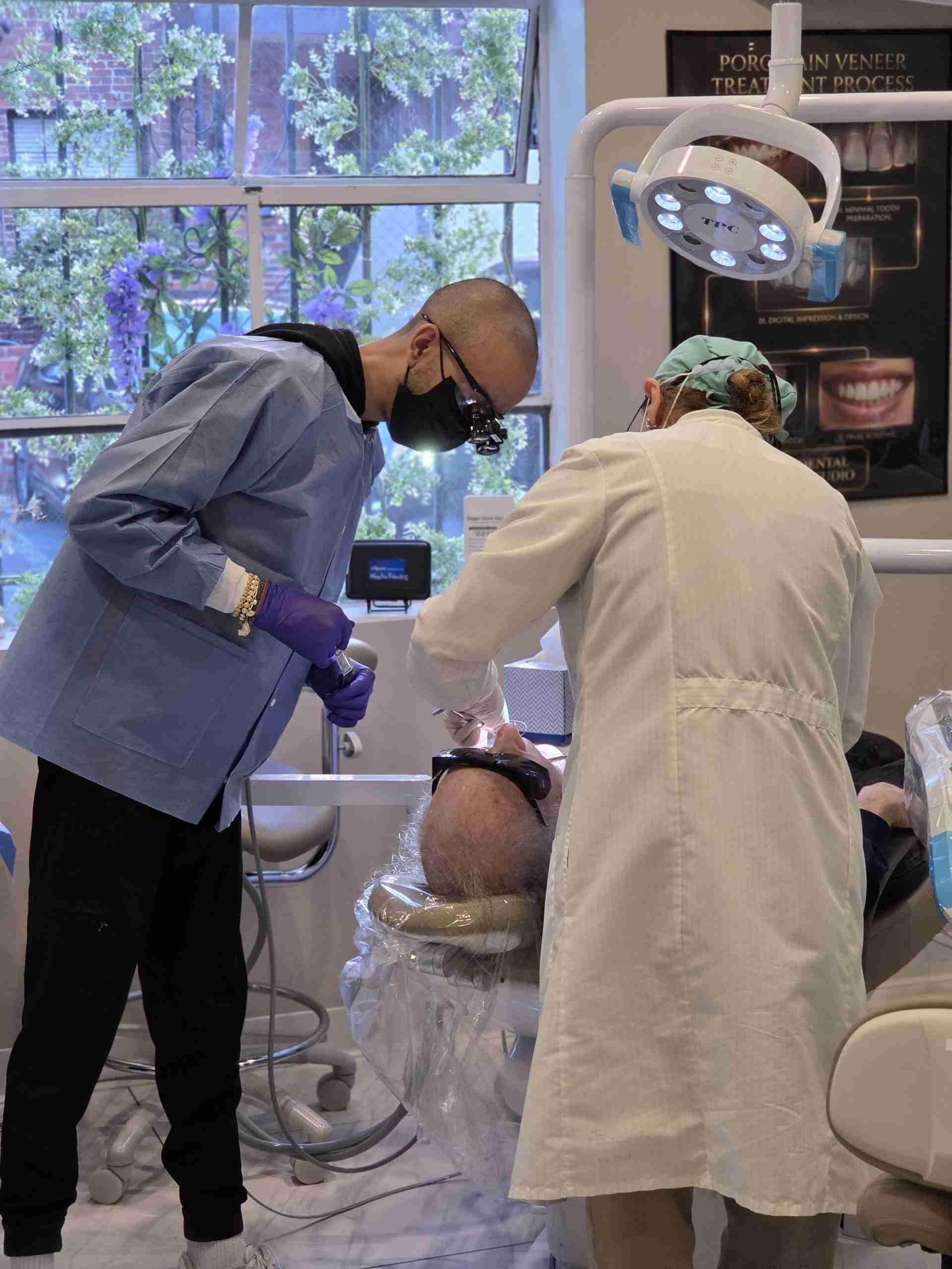
TOLD YOU DO NOT HAVE ENOUGH BONE?
Bone assessments made from a flat X-ray can only approximate height, not width, and width is frequently the deciding dimension. A meaningful number of patients turned down for implants elsewhere turn out to be candidates once the site is measured properly in three dimensions. A scan and an assessment take one appointment. Book with Dr. Shir.
FREQUENTLY ASKED QUESTIONS ABOUT 3D CT SCANS
The questions patients ask most often about cone beam imaging.
How much radiation does a cone beam scan use?
More than a standard dental X-ray and considerably less than a medical CT scan of the head. It is not a zero-risk procedure and we would not describe it as one. It is taken only where the clinical situation genuinely requires three-dimensional information, protective shielding is used, and we will explain what we are looking for before taking it.
What does the scan feel like?
Nothing. You sit or stand still while the machine rotates around your head, which takes under a minute. There is no enclosed tube, nothing touches you, and there is no noise beyond a quiet hum. Patients who find medical CT or MRI difficult generally have no problem with this.
Why do I need a 3D scan when my old dentist only took X-rays?
Because a flat image cannot measure bone width, cannot show exactly where the nerve canal runs relative to a root, and cannot reveal a canal hidden behind another one. For implants, wisdom teeth and complex root canals, that information changes the plan rather than just confirming it. For routine checkups, standard X-rays remain entirely appropriate.
Is the scan covered by insurance?
Coverage varies considerably and some plans treat cone beam imaging differently from standard X-rays or exclude it. Where the scan is part of a larger treatment plan, it is quoted within that plan so you can see the whole figure rather than a series of separate charges. You receive that in writing before anything begins.