

Digital X-Rays in Beverly Hills
Dental Technology
Imaging at a considerably lower radiation dose than film, appearing on screen instantly. This is how decay between teeth is found while it is still a filling.
Seeing What an Examination Cannot
A visual examination shows the surfaces of your teeth. It cannot show what is happening between them, beneath existing fillings, at the root tips or in the bone supporting them, and that is where a great deal of dental disease begins. Digital X-rays use an electronic sensor instead of film, producing an image on screen within seconds at a considerably lower radiation dose. The image can be enlarged, adjusted for contrast and compared directly against your previous ones, which is how small changes get spotted before they become symptoms. Nothing is developed in chemicals and nothing is stored in an envelope.
WHAT DIGITAL X-RAYS SHOW US
What imaging reveals that an examination cannot, and how we use it.

Decay Between Teeth
The contact point where two teeth touch is invisible to the eye and unreachable by a probe. It is one of the most common places for decay to start, and by the time it is visible without imaging it is usually well established.
Decay Beneath Existing Fillings
Old restorations fail quietly at the margins, and new decay develops underneath while the filling still looks intact from above. Imaging is the only way to find that before the tooth starts hurting.
Bone Levels and Gum Disease
Periodontal disease destroys the bone supporting your teeth, and X-rays show that bone level directly. Comparing today's image against one from three years ago turns a subjective impression into a measurement.
Infection at the Root Tip
An abscess forming at the end of a root shows as a dark area on the image, often before there is any pain at all. Finding it early is frequently the difference between a root canal and losing the tooth.
Cracks, Fractures and Trauma
Where a tooth has been knocked or is cracking under a heavy bite, imaging shows the extent below the gum line, which determines whether the tooth is restorable.
Impacted and Unerupted Teeth
Wisdom teeth and, in children, permanent teeth developing beneath the gum are tracked on imaging so problems can be anticipated rather than discovered once they cause pain.
Lower Dose Than Film
Digital sensors need considerably less radiation than the film systems they replaced, and the dose from a routine set of images is very small. Protective shielding is used as standard.
Taken at Intervals Matched to Your Risk
Imaging is not taken automatically at every visit. How often depends on your decay rate, gum health and existing restorations. Some patients need images annually, others less frequently, and we will explain which applies to you.
You See Them on Screen
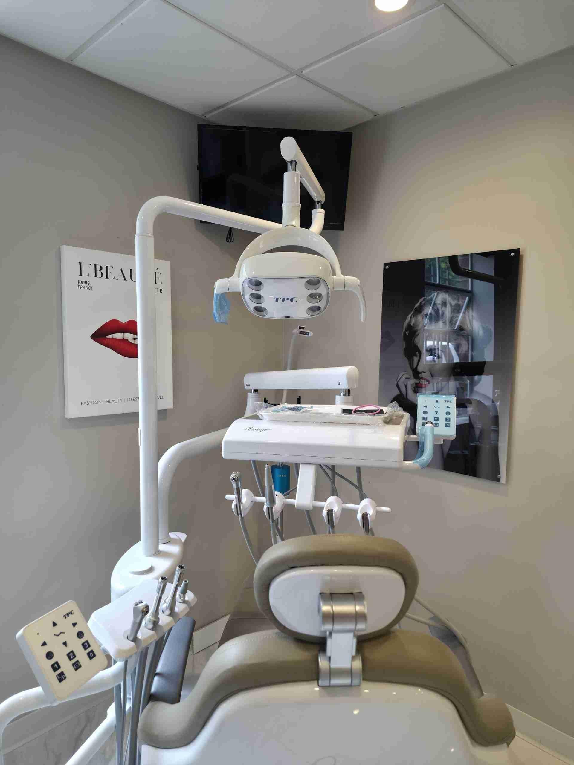
Images appear instantly on a monitor in the room and can be enlarged. Being shown the shadow of decay between your own teeth is considerably more useful than being told it is there.
DIGITAL X-RAYS AND CONVENTIONAL FILM
Both produce a dental image. Almost everything else about them differs.
DIGITAL X-RAYS:
A considerably lower radiation dose, an image on screen within seconds, and the ability to enlarge, adjust contrast and compare against your previous images side by side. Files are stored digitally, shared instantly with a specialist if needed, and never lost in an envelope. No chemicals and no developing.
CONVENTIONAL FILM:
A higher radiation dose, several minutes of chemical developing before anyone can look at the result, and a fixed image that cannot be adjusted. Comparing against past images means finding the physical films. Retakes mean repeating the exposure. There is no clinical advantage that would justify continuing with it.
HOW WE USE IMAGING HERE
The value of imaging is not in taking it. It is in what gets noticed and what gets acted on.

Lower Dose as Standard
Digital sensors throughout, protective shielding used every time, and the dose from a routine set kept very small.

Taken Only When Indicated
Imaging intervals are set by your actual risk rather than taken automatically at every appointment. We will tell you why an image is being taken.

Compared Against Your History
Your previous images are on file and reviewed alongside new ones. Change over time is what matters, and it is invisible if you only ever look at today.

Shown to You, Not Just Described
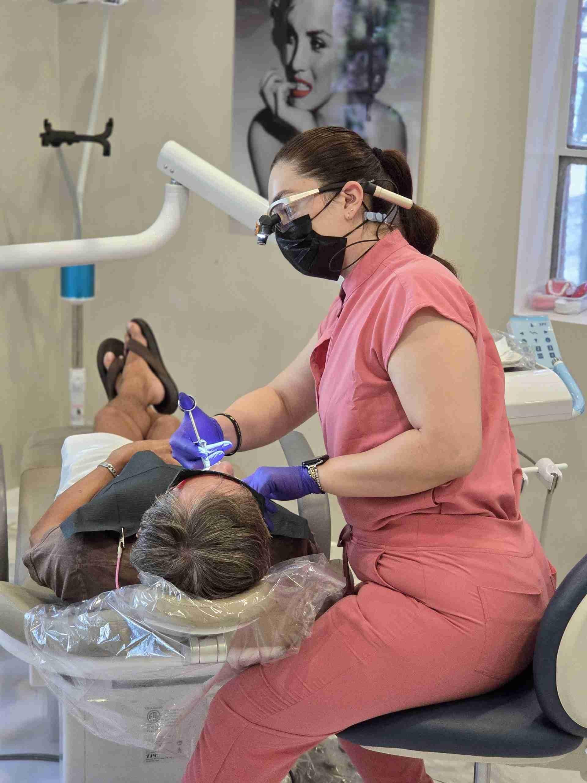
Images go up on the screen in the room. You see the decay, the bone level or the crack yourself before deciding anything.
Lower Dose as Standard
Digital sensors throughout, protective shielding used every time, and the dose from a routine set kept very small.
Taken Only When Indicated
Imaging intervals are set by your actual risk rather than taken automatically at every appointment. We will tell you why an image is being taken.
Compared Against Your History
Your previous images are on file and reviewed alongside new ones. Change over time is what matters, and it is invisible if you only ever look at today.
Shown to You, Not Just Described
Images go up on the screen in the room. You see the decay, the bone level or the crack yourself before deciding anything.
MOST DENTAL PROBLEMS START WHERE YOU CANNOT SEE THEM
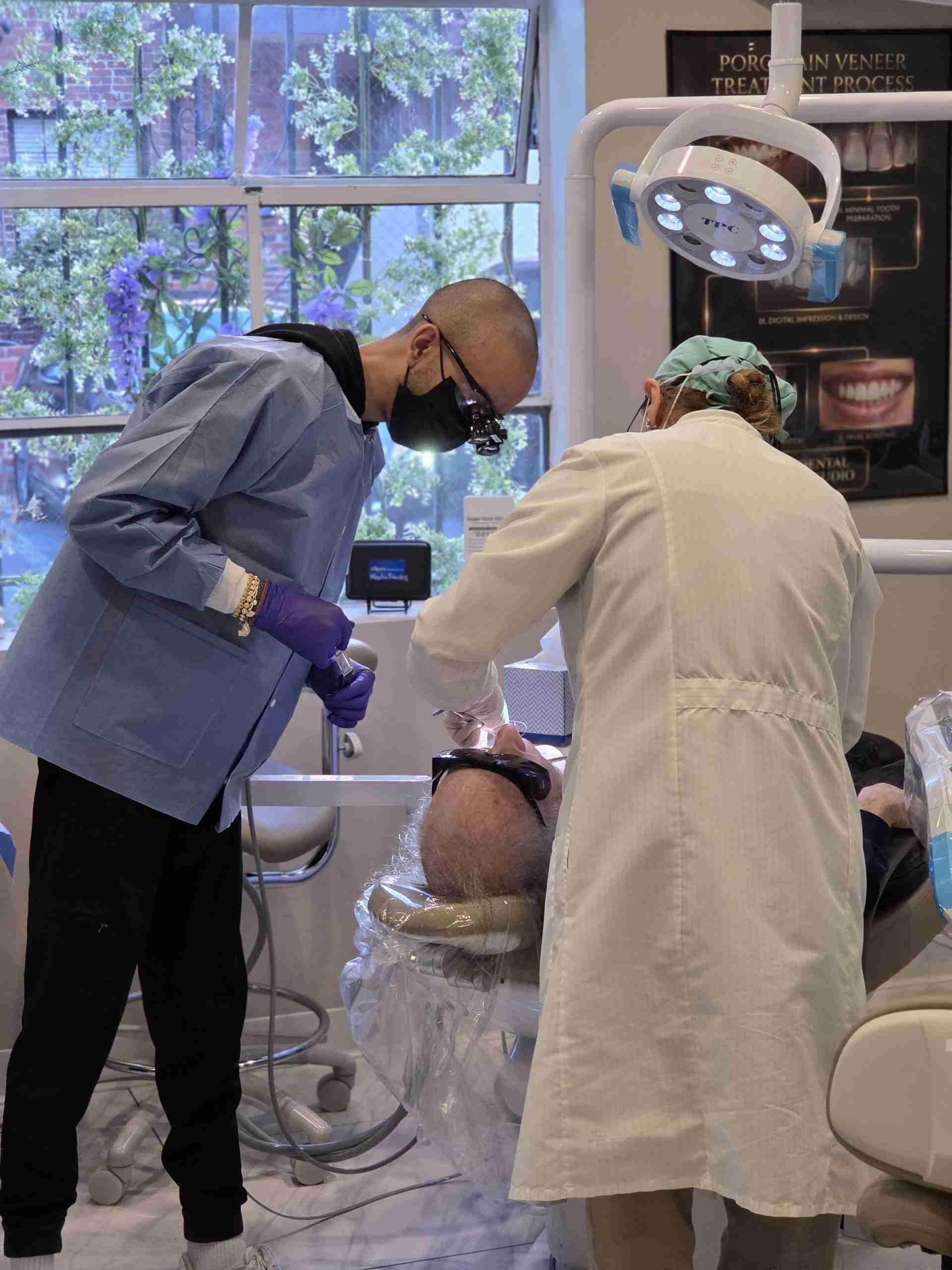
Decay between teeth, failing margins under old fillings and bone loss from gum disease all develop silently, and all of them are visible on imaging long before they produce symptoms. If it has been a while since you had X-rays taken, an examination with current imaging will tell you exactly where you stand. Book with Dr. Shir.
FREQUENTLY ASKED QUESTIONS ABOUT DIGITAL X-RAYS
The questions patients ask most often about dental imaging.
Are dental X-rays safe?
Digital X-rays use a considerably lower radiation dose than the film systems they replaced, and the dose from a routine set is very small. Protective shielding is used as standard, and images are taken at intervals matched to your risk rather than automatically. As with any imaging, the reason for taking it should always be a clinical one, and we will explain what we are looking for.
How often do I need dental X-rays?
It depends on your risk rather than on a fixed schedule. Patients with a history of frequent decay, active gum disease or extensive restorative work generally need imaging more often. Patients with consistently healthy mouths and no restorations need it less. We will tell you what your interval should be and why, rather than taking images by default.
Can I have X-rays if I am pregnant?
Tell us if you are pregnant or think you might be. Routine imaging is usually deferred until after the pregnancy, though where there is an urgent clinical need such as suspected infection, imaging with proper shielding may still be appropriate. That is a conversation we will have with you rather than a decision made without you.
Why do I need X-rays if nothing hurts?
Because most dental disease is painless until it is advanced. Decay between teeth, failing margins under old fillings and bone loss from gum disease all develop silently, and pain generally arrives once the nerve is involved or the tooth is well beyond a simple repair. Imaging finds those things while the treatment is still small.